Mojo presented to the clinic with a wobbly, uncoordinated hind limb gait which was rapidly getting worse. An uncoordinated hind limb gait is often one of the first clinic signs associated with a problem in the cervical (neck area) of the spinal column. The changes in Mojo’s pattern of walking were caused by a loss of proprioception in his hind limbs. Proprioception is the sense which the mammalian body uses to identify the position of neighbouring parts of the body and strength of effort being employed in their movement. Mojo simply didn’t know where his legs were in space as he moved them and this caused him to have an uncoordinated hind limb movement.
Proprioception is often lost in association with changes to the spinal cord, most commonly progressive compression of the cord causing damage to the nerve fibres that carry the information relating to proprioception. An MRI scan was carried out to examine Mojo’s spinal cord for evidence of pathology which may have been caused his loss of proprioception. As soon as the scans appeared it was clear what was causing Mojo’s problems; he has three discs in his neck causing compression of his spinal cord.
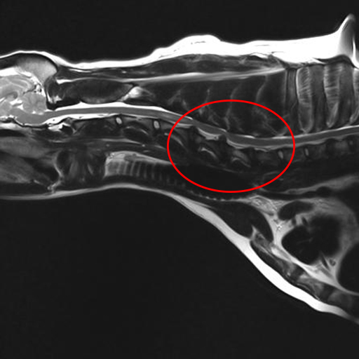
MRI image of Mojo’s neck – circle highlights the discs compressing the spinal cord.
Mojo’s owners were faced with the choice of allowing his disease process to take its course – likely resulting in progressive paralysis of his back legs and subsequent euthanasia – or to commit to a surgical intervention to try and stop the progression of disease. Mojo’s owners made the commitment to put their beloved pet through surgery to try and give him back the life he so adored!
Surgery of the spinal cord isn’t without risk as there are a multitude of vital structures in the neck, damage to any one of which could have devastating effects for the patients – something that is clear during tonight’s episode! Mojo was prepared for theatre and surgery was carried to fuse Mojo’s neck to resolve the compression of his spinal cord. Specially designed bolts were placed between the bones of his cervical spine to push them apart and reduce the compression of the cord. The bones of the spine were then fused together using a combination of plates, screws and bone marrow.
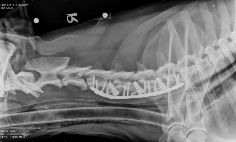
Post-surgery radiograph showing bolts, screws and plates stabilising Mojo’s neck.
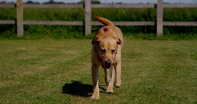
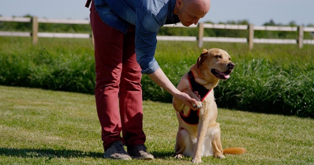
After spinal surgery it is not unusual for patients to be unable to walk for a day or two as the spinal cord recovers. Rehabilitation started immediately after Mojo’s surgery with the physiotherapy team carrying our exercises to try and improve his proprioception and muscle strength. It was to jubilation in kennels that Mojo took his first steps only 72 hours after surgery and once he was up and about there was no holding him back. Mojo recovered at an exponential rate and the proprioceptive deficits in his hind limbs rapidly improved.
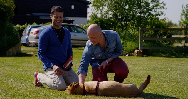
Mojo is a much loved member of his family and we understand fully the emotions owners go through when making the decision to put a family member through surgery – it is our job to hold owners hands through this journey.
In a case like Mojo’s the only way we can achieve a positive surgical outcome is through team work. The skills of a diverse care giving team comprising nurses, vets, animal care assistants, hydrotherapists and physiotherapists are all called upon to bring patients through surgeries such as spinal fusions and it is the skill and dedication of member of the team that contributes to the positive outcomes we achieve. Mojo will undergo regular check-ups throughout his life and we will keep you updated on his progress.

1
2

3
4
5